Image

Imaging the cerebral reality of long COVID symptoms? Researchers at Institut Fresnel (IMOTHEP team) have done just that at CERIMED (2 Carnot STAR units), in collaboration with IHU Méditerranée Infection.
Tuesday, January 19, 2021
Imaging the cerebral reality of long COVID symptoms? Researchers at Institut Fresnel (IMOTHEP team) have done just that at CERIMED (2 Carnot STAR units), in collaboration with IHU Méditerranée Infection.
The metabolic PET* imaging study - carried out on 35 patients with a biologically confirmed diagnosis of COVID19 infection and persistent functional complaints at least 3 weeks after the initial episode (3 months on average, and up to 5 months), compared with 44 healthy uninfected subjects - has been published in the European Journal of Nuclear Medicine & Molecular Imaging.
The results of this study confirm patients' feelings & could "suggest further studies to demonstrate the value of early treatment of infection and inflammation of the ENT sphere, in order to avoid possible secondary extension of the pathological process to the brain. In the absence of sequellar structural lesions demonstrated at this stage in morphological imaging (notably MRI), these results could also suggest approaches to reactivating this hypofunctional cerebral network through sensory, cognitive and physical re-education and rehabilitation strategies."
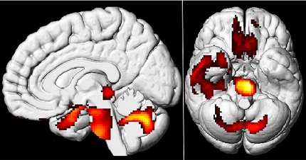
Figure: Cerebral hypometabolism found on 18F-FDG PET in patients with long COVID. Compared with healthy subjects with no history of COVID19 infection, patients with long COVID showed hypometabolism of the olfactory bulb and connected limbic regions, extending to the brainstem and cerebellum.